What Keeps the Heart Beating Without Ever Stopping to Rest?
Every other muscle in the human body eventually gives out. Run up a few flights of stairs and your legs will burn. Grip a pull-up bar too long and your forearms will fail. Even the diaffragm, which works constantly to keep you breathing, gets brief pauses between breaths. The heart gets none of that. From roughly three weeks after conception until the moment of death, it contracts, relaxes, and contracts again without a single beat of true rest.
That fact is strange enough to deserve an explanation. Skeletal muscle fatigues because of chemical and structural limits: it burns through energy reserves, accumulates waste products, and depends on nerve signals that can tire. If the heart were built the same way, a lifetime of continuous contraction would be physically impossible. Instead, cardiac muscle is built on a completely different design, one that solves the problem of endurance at the level of individual cells, electrical wiring, and energy supply.
Understanding how the heart manages this is really an answer to a bigger question: what does true, uninterrupted reliability require, and how does biology build something that cannot afford to fail even once?
A Muscle Unlike Any Other in the Body
Skeletal muscle, the kind that moves your arms and legs, is designed for power and control, not endurance. Its fibers are long, arranged in parallel bundles, and built to contract briefly and forcefully before resting. Cardiac muscle looks and behaves differently from the moment you examine it under a microscope.
Heart muscle cells, called cardiomyocytes, are shorter and branched, connecting to one another in a dense, interlocking network. At the junctions between these cells sit structures called intercalated discs, which contain two critical features: mechanical anchors that let the cells pull together as a single unit, and electrical channels called gap junctions that let electrical signals pass instantly from one cell to the next. The result is that the heart doesn’t behave like a bundle of separate fibers. It behaves like one continuous, coordinated sheet of tissue, so that a signal starting in one corner spreads through the entire chamber in a fraction of a second.
Cardiac cells are also packed with far more mitochondria than skeletal muscle, often filling a third or more of the cell’s volume. Mitochondria are the structures that convert oxygen and nutrients into usable energy, and this density reflects the heart’s central problem: it needs a constant, uninterrupted fuel supply, because unlike skeletal muscle, it has no real opportunity to catch up later.
The Heart Generates Its Own Rhythm
Perhaps the most important fact about the heart is one that surprises most people: it does not wait for instructions from the brain to beat. It generates its own electrical signal, a property called autorhythmicity, or automaticity.
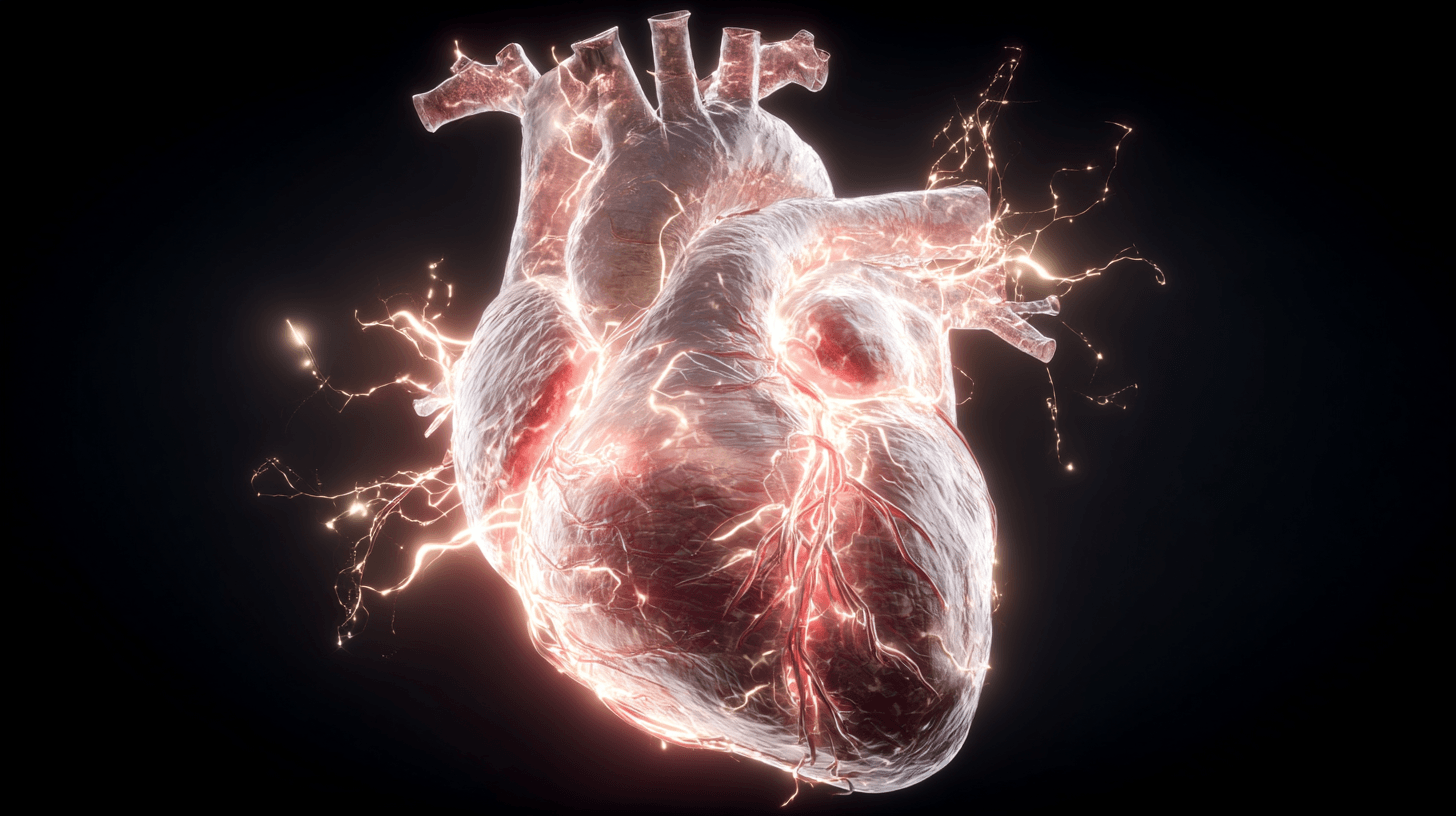
Inside the wall of the right atrium sits a small cluster of specialized cells known as the sinoatrial node, or SA node. These cells are unusual because their membranes are naturally unstable. Rather than sitting quietly until stimulated, ions leak across the membrane on their own, causing the cell’s electrical charge to drift upward until it crosses a threshold and fires, all without any external trigger. Once it fires, the signal spreads across both atria, causing them to contract, and then converges on a second cluster of cells called the atrioventricular node, or AV node.
The AV node briefly delays the signal, allowing the atria to finish emptying blood into the ventricles below before the ventricles contract. The signal then races down a pathway called the bundle of His and out through branching fibers called Purkinje fibers, which spread the impulse through the thick ventricular walls almost simultaneously. This entire sequence, from the first spark in the SA node to the final contraction of the ventricles, takes well under a second.
Because the SA node fires spontaneously and repeatedly, the heart doesn’t need a continuous stream of commands. It needs only oversight. This is a crucial distinction: the nervous system adjusts the heart’s pace, but it does not create the heartbeat itself. That job belongs entirely to the heart’s own tissue.
Why the Heart Can Beat in Isolation
The clearest proof of the heart’s self-generated rhythm comes from an unusual but well-documented fact: a heart removed from the body, and completely disconnected from the brain and nervous system, will keep beating on its own for a period of time if it is kept oxygenated and warm. This is not a hypothetical claim. It is the basis of heart transplantation.
When surgeons transplant a heart, they do not reconnect the donor heart’s nerves to the recipient’s nervous system. There is currently no reliable surgical technique for doing so. The transplanted heart is denervated, cut off from direct neural control, yet it beats steadily for the rest of the recipient’s life, adjusting its rate more slowly through hormones circulating in the blood rather than through nerve signals. This tells researchers something important: the heartbeat is not fundamentally a product of brain activity. It is a property of the heart’s own tissue, and the nervous system’s role is closer to that of a governor than an engine.
The Problem of Fatigue, and How the Heart Avoids It
Skeletal muscle fatigues for several overlapping reasons: it depletes stored energy, allows waste products like lactate to accumulate, and can enter a sustained, rigid contraction called tetanus if stimulated rapidly enough. Cardiac muscle is structurally protected against all three problems.
The most important safeguard involves timing. After a cardiac cell contracts, it enters a period called the absolute refractory period, during which it cannot be re-stimulated no matter how strong the incoming signal is. This window lasts almost as long as the contraction itself, which means the heart muscle is guaranteed a brief pause before it can fire again. Skeletal muscle has a much shorter refractory period, which allows it to be driven into tetanus, a sustained, non-relaxing contraction, if it is stimulated fast enough. The heart’s long refractory period makes this kind of sustained contraction physically impossible, because by the time a new electrical signal could arrive, the previous contraction has already finished relaxing. This is not an incidental detail. It is the reason the heart can pump at all. If cardiac muscle could tetanize the way skeletal muscle does, the heart would lock into a single contraction and stop moving blood entirely.
The second safeguard is fuel supply. Skeletal muscle can rely partly on quick, inefficient energy pathways that generate energy without oxygen, which is exactly what allows a sprinter to keep running even as oxygen debt builds up. The heart relies almost entirely on oxygen-based metabolism, and it needs a large, unceasing blood supply of its own to make that possible. This is delivered through the coronary arteries, which branch across the heart’s surface and feed its muscle directly. When one of these arteries becomes blocked, cardiac tissue downstream is quickly starved of oxygen, which is what causes a heart attack, or myocardial infarction. The heart’s dependence on continuous oxygen delivery is the cost of its continuous performance: it cannot store enough energy to skip even a few minutes of blood flow.
The Nervous System Adjusts the Pace, Not the Existence, of the Heartbeat
Although the heart generates its own rhythm, it does not operate in isolation from the rest of the body. The autonomic nervous system, the branch responsible for automatic, non-conscious regulation, adjusts the heart’s rate to match the body’s needs.
The sympathetic nervous system, associated with stress, exercise, and the so-called fight-or-flight response, releases signals that speed the SA node’s firing rate and strengthen each contraction. The parasympathetic nervous system, primarily through a large nerve called the vagus nerve, slows the SA node down during rest. Most people’s resting heart rate is well below the SA node’s natural, undisturbed firing rate, largely because the vagus nerve is constantly applying a mild brake. This is why certain medical procedures or nerve injuries that affect vagal tone can noticeably speed up a resting heart rate.
Hormones add another layer of adjustment. Adrenaline and noradrenaline, released during moments of exertion or stress, bind to receptors on cardiac cells and increase both heart rate and the force of each contraction. This is one reason the transplanted, denervated heart described earlier can still respond to exercise: circulating hormones reach it even without direct nerve connections, just more gradually than a fully innervated heart would respond.
What Happens When the System Breaks Down
The heart’s rhythm is remarkably stable, but it is not indestructible, and understanding its failures illuminates how the healthy system works. If the SA node is damaged or its signal is blocked before reaching the ventricles, a condition called heart block, backup pacemaker tissue elsewhere in the heart can take over, though usually at a slower and less coordinated rate. This is one reason autorhythmicity exists at multiple points in the heart’s conduction system, not just the SA node. It provides redundancy.
When the heart’s electrical activity becomes chaotic rather than coordinated, a condition called fibrillation, the muscle fibers contract independently rather than as a unit, and the heart loses its ability to pump blood effectively. Atrial fibrillation is uncomfortable but often survivable for extended periods; ventricular fibrillation is immediately life-threatening, because the ventricles are responsible for pushing blood to the lungs and the rest of the body. This is the condition that defibrillators are designed to correct, delivering a controlled electrical shock intended to reset the heart’s cells simultaneously so that the SA node can resume normal control.
Artificial pacemakers, first implanted successfully in the late 1950s, work by mimicking the SA node’s function when the heart’s natural pacing system fails or becomes unreliable. They do not replace the heart’s ability to contract, only its ability to initiate a regular signal, which is a useful illustration of how separable the heart’s mechanical and electrical functions really are.
A Design Built Entirely Around Reliability
Nearly every structural feature of the heart, from its branching, electrically connected cells to its long refractory period to its dense mitochondrial supply, exists to solve the same underlying problem: how do you build a pump that can never afford to stop, even briefly, for the entire span of a human life. Skeletal muscle can rest because the body can tolerate uneven movement. The circulatory system cannot tolerate uneven pumping, because organs throughout the body depend on a continuous supply of oxygenated blood.
The heart’s autorhythmicity means it does not depend on a fragile, centralized decision-maker to keep functioning. Its refractory period makes sustained, catastrophic contraction physically impossible. Its dense energy supply reflects a system built entirely around the assumption that pausing is not an option.
A resting adult heart beats roughly 60 to 100 times per minute, which adds up to something in the range of 2.5 to 3.5 billion beats over an average lifetime, depending on individual heart rate and lifespan. No other muscle in the body is asked to perform anything close to that number of contractions without a single true rest. The heart manages it not through extraordinary willpower or unusual toughness, but through a specific, elegant biological architecture: a rhythm it generates itself, a structural safeguard that makes early re-firing impossible, and a fuel supply built to match the relentless nature of its own workload.
Frequently Asked Questions
Does the brain control every individual heartbeat?
No. The heart’s own tissue, primarily the sinoatrial node, generates each heartbeat automatically. The nervous system adjusts the speed and strength of these beats but does not create them.
Can the heart really keep beating outside the body?
Yes, for a limited time, if it is kept warm and supplied with oxygen and nutrients. This principle underlies heart transplantation and certain heart-preservation techniques used before transplant surgery.
Why doesn’t the heart get sore the way other muscles do?
Cardiac muscle has a long refractory period that prevents the sustained, locked contractions that cause other muscles to fatigue, along with an unusually dense supply of energy-producing mitochondria and continuous blood flow through the coronary arteries.
What causes a heart attack if the heart is so resilient?
A heart attack occurs when a coronary artery becomes blocked, cutting off oxygen to part of the heart muscle. Because cardiac tissue depends almost entirely on a steady oxygen supply, even a short interruption can cause lasting damage.
How many times does the heart beat in a lifetime?
Estimates vary with individual heart rate and lifespan, but a commonly cited range is approximately 2.5 to 3.5 billion beats over an average human life.